In the early days of cancer research, we relied on surgical microscopes, stained slides, and educated guesses. Now, we’re seeing tumor biology unfold on molecular levels that used to be pure theory. And while treatments are slowly catching up, cancer detection and mapping are evolving at a pace that feels—finally—appropriate to the urgency of the disease. The technology isn’t just advanced. It’s reshaping how we even define what a tumor is, what it does, and how it communicates with the rest of the body.
This isn’t about one miracle drug or some flashy device. It’s the cumulative progress of better imaging, deeper gene profiling, smarter delivery systems, and most importantly, more specific targeting of what actually drives malignancy. The recent breakthroughs aren’t curing cancer, but they’re doing something arguably just as important: narrowing the blind spots.
Beyond Imaging: The Race Toward Live Tumor Profiling
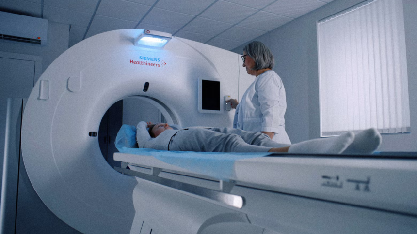
Traditional imaging has always had limits. MRI and CT scans can show size and location, but not activity. PET scans can indicate metabolism but often miss nuance. That’s changing as molecular imaging becomes more refined and context-aware. We’re no longer just looking at masses. We’re looking at real-time biochemical behavior.
One of the most promising paths is metabolic flux imaging, which captures how tumor cells process energy and build biomass. These aren’t snapshots; they’re live feedback loops of cellular decision-making. Researchers at several institutions are using hyperpolarized MRI agents that make it possible to watch pyruvate conversion in real time—a powerful indicator of tumor aggressiveness.
This has implications far beyond the lab. With early metabolic mapping, oncologists can make faster decisions about whether a tumor is worth biopsying or treating aggressively. That’s especially important in areas like prostate or thyroid cancers, where the distinction between dormant and aggressive disease can mean the difference between watchful waiting and invasive therapy.
A Cellular Whisper Network: Tumor Microenvironment Decoded
Tumors don’t exist in isolation. They’re shaped by, and reshape, their microenvironment—fibroblasts, immune cells, vasculature, and more. A tumor’s survival depends as much on its neighborhood as on its internal mutations. And until recently, we didn’t have the tools to break down those interactions at high resolution.
Single-cell RNA sequencing has pushed this field into overdrive. We can now identify not just what genes are expressed in a sample, but exactly which cell types are saying what. Even better, we can locate them spatially. Spatial transcriptomics, a newer technique, overlays gene activity data directly onto tissue architecture. The result? We’re mapping not just what cells are doing but where they’re doing it.
That’s how we landed on one of the most promising clues in years: protein discovery in disease-associated locations within tumors that had previously looked benign under standard pathology. This wasn’t a matter of a single errant cell type. It was a quiet, coordinated shift in signaling proteins expressed at the tumor’s borders—clear evidence that microenvironmental crosstalk was facilitating spread, even before the tumor itself looked aggressive.
That kind of early molecular signature opens up an entirely new layer of diagnostics. It’s not about what cancer is. It’s about what it’s preparing to do.
Liquid Biopsies Are Finally Growing Up
For years, liquid biopsies sounded like science fiction: a vial of blood replacing a tissue biopsy, catching cancers in their earliest stages, tracking treatment in real time. The hype got ahead of the data. But that’s finally starting to shift.
Newer assays are far more sensitive to circulating tumor DNA (ctDNA), and crucially, they’re getting better at distinguishing it from background noise. False positives were once the Achilles heel of early-stage detection; newer models use machine learning to filter out unrelated DNA fragments and home in on mutations with high oncogenic potential.
Some of the most useful advances involve methylation patterning. Methylation signatures aren’t just tumor-specific—they’re tissue-specific. That means we’re not just detecting cancer in the blood; we’re narrowing down where it’s likely to be forming. For high-risk populations, this could mean truly preventive interventions.
Add in exosome analysis—where tumor-derived vesicles are isolated from blood or saliva—and the picture gets sharper. Exosomes carry genetic material, proteins, and signaling molecules. In some patients, their presence can reveal recurrence before any clinical symptoms appear.
Precision Therapies That Actually Earn the Label
Targeted therapies aren’t new. But most have been… generously labeled. “Targeted” often meant a drug inhibited a receptor that the tumor sometimes overexpressed. The reality was closer to selective toxicity than true targeting. That’s slowly changing as therapies are built on detailed knowledge of molecular drivers.
Antibody-drug conjugates (ADCs) are a great example. They link a cytotoxic drug to an antibody that binds a specific tumor antigen, releasing the drug only when the antibody hits its target. That’s been done before, but newer ADCs have smarter linkers—pH-sensitive or enzymatically cleaved—that reduce off-target spill.
Meanwhile, bispecific T-cell engagers (BiTEs) are creating new ways to redirect the immune system. They bind to a tumor antigen on one end and to CD3 on a T-cell on the other, drawing immune cells into proximity with tumor cells even in “cold” tumors that previously resisted checkpoint inhibitors.
These therapies are being guided not just by tissue biopsies, but by evolving data from blood, imaging, and sometimes wearable biosensors. In the case of brain injuries in veterans or patients with neuro-oncology crossover diagnoses, this multi-modal approach becomes the difference between finding a usable treatment and missing a diagnosis entirely.
The AI You’re Not Hearing About Yet
Most of the headlines about AI in oncology are stuck on radiology. But the more interesting applications are happening behind the curtain—in drug discovery, in predictive modeling, and in cross-referencing genomic data that’s far too vast for humans to parse.
Some of the newer models are focused entirely on discovering overlooked driver mutations. These aren’t the high-profile ones like KRAS or EGFR, but the ones buried in less-studied exons, or the ones that only matter when combined with a particular microenvironmental signal. Others are being trained to simulate how a tumor will evolve under treatment pressure—so oncologists can strategize a few moves ahead, not just react to resistance.
Then there’s the use of AI in clinical trials. Matching patients to trials based on genetics and tumor markers is notoriously clunky, but newer systems are using natural language processing to crawl both structured and unstructured data and find candidates who would’ve never been flagged by traditional metrics. That means faster enrollment, more representative samples, and a shot at more meaningful results.
Closing Note: Where the Work Gets Interesting
The hardest part of cancer research isn’t the science. It’s the translation. Bridging what we now understand on a molecular level with what’s available to patients in community clinics still lags. But that gap is shrinking. These new tools aren’t just collecting data—they’re informing real decisions, in real time.
Cancer’s biggest trick has always been its variability. It mutates. It adapts. It hides behind healthy tissue or blends into normal variation. But the technologies surfacing now are meeting that challenge with specificity and speed that wasn’t possible even five years ago.
There’s still no silver bullet. But there are now hundreds of silver scalpels—quiet, precise, and ready to cut through the noise.
Lynn Martelli is an editor at Readability. She received her MFA in Creative Writing from Antioch University and has worked as an editor for over 10 years. Lynn has edited a wide variety of books, including fiction, non-fiction, memoirs, and more. In her free time, Lynn enjoys reading, writing, and spending time with her family and friends.