

Key Takeaways
SPECT stands for single-photon emission computed tomography. A brain SPECT scan uses an injected radiotracer and a gamma camera to create images reflecting regional blood flow at the time the tracer is taken up by the brain.
The resulting images can show areas with relatively higher or lower perfusion, but a perfusion pattern does not automatically identify its cause.
Brain SPECT has established and developing applications in certain neurological settings. Its use for diagnosing individual psychiatric disorders remains debated and is not accepted as a standalone diagnostic method.
Amen Clinics incorporates SPECT imaging into some comprehensive evaluations alongside clinical history, interviews, questionnaires, cognitive testing, and other information.
Scan findings should be interpreted in the context of symptoms, medications, substance use, neurological history, previous injuries, and other medical information.
SPECT involves exposure to ionizing radiation. Although the amount used for diagnostic imaging is generally limited, patients should discuss pregnancy, breastfeeding, medications, allergies, and previous imaging with the clinical team.
Patients should ask how the scan could affect their evaluation, what alternatives are available, who will interpret the images, and whether the expected information justifies the cost and radiation exposure.
The Science Behind SPECT Scans: Understanding Brain Perfusion Imaging
How SPECT Imaging Works
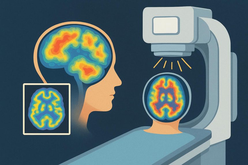
A brain SPECT scan is a nuclear medicine procedure. It begins with the administration of a radiopharmaceutical, commonly called a radiotracer. The tracer travels through the bloodstream and distributes within the brain according to regional blood flow.
After an uptake period, the patient lies on a table while a gamma camera rotates around the head. The camera detects energy emitted by the radiotracer. A computer then reconstructs that information into three-dimensional images.
The image reflects the distribution of the tracer during a particular period. It does not continuously monitor brain activity in real time. What a person is doing around the time of the injection can affect the resulting perfusion pattern, which is why the clinical team provides specific instructions about the environment and activity.
Amen Clinics states that a typical evaluation may include two brain SPECT studies. One is performed in connection with a concentration task, while the other examines the brain in a resting condition. The precise protocol depends on the evaluation ordered for the patient.
A consistent setting is important. Noise, conversation, movement, medication, caffeine, alcohol, and other substances may affect cerebral blood flow. Patients should follow the instructions provided by the imaging facility and should not independently stop prescribed medication.
What Brain SPECT Images Can Show
Brain SPECT primarily provides information about regional cerebral perfusion. The images may show areas receiving relatively more or less blood flow compared with other regions or reference data.
This information is different from a routine structural MRI or CT scan. CT and conventional MRI are commonly used to examine anatomy, such as bleeding, masses, tissue injury, or other structural changes. SPECT is used to evaluate aspects of function through radiotracer distribution.
SPECT findings are not unique to one condition. A region of relatively increased or decreased perfusion may be influenced by several factors, including:
- Neurological disease
- Previous brain injury
- Seizure activity
- Medication
- Substance use
- Sleep
- Pain
- Movement during the procedure
- The patient’s activity during tracer uptake
- Technical and interpretive variables
For this reason, an image cannot be translated directly into a conclusion such as “this pattern proves ADHD” or “this region confirms anxiety.” Symptoms that appear similar can have different causes, and different people with the same diagnosis may not display identical imaging findings.
Appropriate interpretation requires knowledge of the scan protocol, clinical question, patient history, medications, and other test results. When structural disease is suspected, an MRI, CT scan, laboratory test, or another evaluation may still be necessary.
SPECT Imaging Within an Amen Clinics Evaluation
How Imaging May Contribute to an Individualized Assessment
Amen Clinics describes brain SPECT as one component of its broader evaluation process. The clinic states that its clinicians review brain perfusion images alongside clinical interviews, personal and family history, questionnaires, cognitive testing, and other information.
People considering a spect brain scan at Amen Clinics should understand how the clinic proposes to use the imaging in their individual case. A useful question is whether the scan is intended to investigate a neurological concern, add context to a complex clinical history, establish a baseline, or serve another defined purpose.
An individualized treatment plan may include psychotherapy, medication, medical referrals, sleep-related recommendations, exercise, nutrition, or other interventions. The presence of an imaging result does not establish that any particular intervention will work.
Treatment decisions should still consider:
- The patient’s symptoms and diagnosis
- Medical and psychiatric history
- Previous treatment responses
- Current medications and supplements
- Possible adverse effects
- Coexisting medical conditions
- Patient preferences
- The quality of evidence supporting the proposed treatment
Patients can ask the clinician to explain which recommendations come directly from established clinical findings and which are influenced by the SPECT interpretation. They can also ask what would be recommended if imaging were not available.
Understanding the Limits of Psychiatric Interpretation
Brain imaging has contributed greatly to research on psychiatric and neurological conditions. Studies have identified average differences among groups of participants with depression, ADHD, anxiety, schizophrenia, and other conditions.
A group-level research finding does not necessarily function as a diagnostic test for one person. There can be substantial overlap between people with and without a particular diagnosis. Different conditions can also produce similar symptoms or imaging findings.
The American Psychiatric Association has reported that brain-imaging biomarkers have not demonstrated sufficient sensitivity and specificity to classify individual psychiatric cases reliably. This does not mean that imaging has no value. It means that its role must be described accurately.
A SPECT scan should not replace a comprehensive psychiatric or medical evaluation. Diagnoses such as depression, anxiety disorders, ADHD, and PTSD are generally based on clinical criteria that consider symptoms, duration, impairment, history, and alternative explanations.
Patients should be cautious about claims that a single brain pattern can:
- Confirm a psychiatric diagnosis
- Reveal the precise cause of a symptom
- Select a medication with certainty
- Predict an individual treatment response
- Guarantee a better outcome
- Demonstrate that an intervention has cured a disorder
Amen Clinics states in its patient FAQ that a SPECT scan does not provide a diagnosis on its own. According to the clinic, diagnoses are developed from multiple sources of information, including clinical history, interviews, questionnaires, imaging, and neuropsychological testing.
Evaluating Evidence, Outcomes, and Treatment Claims
Why Patient Stories Are Not Clinical Proof
Personal accounts can help prospective patients understand what an experience felt like, but they cannot establish whether a scan or treatment will produce the same outcome for someone else.
A patient may receive several interventions after a SPECT evaluation. Medication changes, psychotherapy, improved sleep, reduced substance use, exercise, nutrition, social support, and the passage of time may all influence progress. It can be difficult to determine how much of the change resulted from the imaging itself.
Testimonials also tend to feature people who chose to share their experiences. They may not represent every person who received the service, including those whose symptoms did not improve or who discontinued treatment.
“A compelling patient experience can raise useful questions, but it does not replace controlled evidence about safety, diagnostic accuracy, or treatment effectiveness.”
When reviewing an outcome claim, patients can ask:
- How was improvement defined?
- Was it measured with a validated questionnaire?
- How many patients were included?
- How many completed follow-up?
- Was there a comparison group?
- Were the results independently reviewed?
- Which treatments were provided after imaging?
- Can the findings be applied to patients with different conditions?
A high percentage alone does not answer these questions. Study design, follow-up rates, patient selection, and the way outcomes were measured all affect how a result should be interpreted.
Questions to Ask Before Making a Decision
The decision to undergo SPECT imaging should be based on the individual clinical situation. Patients may want to discuss the procedure with both the clinic offering it and an independent physician who understands their medical history.
Questions worth asking include:
- What specific clinical question is the scan intended to answer?
- Is brain SPECT routinely recommended for my suspected condition?
- What evidence supports using it in my situation?
- Could MRI, CT, EEG, laboratory testing, or neuropsychological assessment provide more appropriate information?
- How will the result change my treatment?
- What happens if the scan is normal or inconclusive?
- Who will interpret the images?
- Will a nuclear medicine physician or radiologist issue a report?
- How much radiation will I receive?
- Will the evaluation involve one scan or two?
- What are the total costs?
- Is the procedure covered by insurance?
- Can I obtain copies of the images and written reports?
Patients should also ask whether recommended treatments are supported by established guidelines for their condition. A scan may provide additional information, but it does not remove the need to assess the evidence, benefits, risks, and alternatives for each treatment.
Seeking a second opinion is reasonable, especially when the proposed evaluation is costly, involves radiation, or may lead to major changes in medication or care.
Your SPECT Appointment: What to Expect at Amen Clinics
The Brain SPECT Process
The exact process varies according to the protocol and type of evaluation. Amen Clinics states that its typical comprehensive approach may involve resting and concentration scans performed on different parts of the evaluation.
A general brain SPECT process may include:
- Pre-scan review: Staff review medical history, medications, supplements, substance use, pregnancy or breastfeeding status, and other factors that could affect safety or interpretation.
- Preparation: The patient follows instructions concerning food, caffeine, alcohol, nicotine, exercise, medication, and arrival time.
- IV placement and tracer injection: A small intravenous line is used to administer the radiotracer.
- Tracer-uptake period: The patient remains in a controlled setting or completes the activity required by the protocol.
- Image acquisition: The patient lies still while the gamma camera rotates around the head and collects information.
- Clinical interpretation: Qualified professionals review the images in connection with the clinical history and other assessment results.
- Results appointment: A clinician explains the findings, their limitations, and any proposed next steps.
Amen Clinics reports that each image-acquisition session generally takes about 20 to 25 minutes. The entire appointment may take longer because of registration, IV placement, uptake time, and other parts of the evaluation.
Movement can reduce image quality. Patients who experience claustrophobia, pain while lying down, difficulty remaining still, or anxiety about needles should tell the clinic before the appointment.
Safety, Results, and Next Steps
SPECT uses ionizing radiation. The exposure is generally limited, but it is not zero. The appropriate dose and expected benefit should be considered for each patient, particularly when two scans or repeat imaging are proposed.
Patients should tell the clinic if they are pregnant, could be pregnant, or are breastfeeding. They should also disclose allergies, kidney or liver concerns, recent nuclear medicine procedures, and all medications and supplements.
Allergic reactions to radiotracers are uncommon, but they can occur. The injection may also cause brief discomfort, bruising, or redness at the IV site. The clinical team should explain the specific radiopharmaceutical, expected dose, precautions, and aftercare instructions.
Results should be explained in plain language. A useful discussion distinguishes among:
- What the scan directly shows
- What the interpreter believes the pattern may suggest
- What cannot be determined from the scan
- How the result compares with the clinical history
- Whether additional testing is recommended
- How the information affects the proposed treatment
Medication changes should be made only with the prescribing clinician. Patients should understand the expected benefits, possible adverse effects, alternatives, and monitoring plan for every recommended treatment.
SPECT imaging may contribute information to a broader evaluation, but it cannot guarantee diagnostic certainty or treatment success. A careful decision considers the clinical purpose, evidence, limitations, radiation exposure, cost, and available alternatives.
FAQ
What mental health conditions can a SPECT scan diagnose?
A SPECT scan cannot independently diagnose anxiety, depression, ADHD, PTSD, or another psychiatric disorder. It shows regional perfusion patterns that must be interpreted with the patient’s clinical history and other assessments. Psychiatric diagnoses generally rely on established clinical criteria.
How does SPECT differ from MRI or CT?
SPECT uses a radiotracer to show aspects of regional blood flow and function. Routine MRI and CT primarily show anatomy and structural abnormalities. Each examination answers different clinical questions, and one does not automatically replace the others.
Can SPECT be used as a standalone diagnostic tool?
No. Amen Clinics states that SPECT alone does not provide a diagnosis. Clinical interviews, history, symptom assessment, cognitive testing, physical or neurological evaluation, and other medical tests may all be relevant.
Who interprets brain SPECT results?
Nuclear medicine imaging is generally interpreted by a physician with appropriate training in nuclear medicine or radiology. Other clinicians may use the report and images as part of a broader evaluation. Patients should ask who will formally interpret their study and what qualifications that person holds.
How long does it take to receive the results?
The schedule varies by clinic, protocol, and evaluation package. Patients should ask when the images will be interpreted, whether they will receive a written report, and when the results consultation will occur.
Are there risks associated with a SPECT scan?
SPECT requires an injected radiotracer and involves ionizing radiation. The radiation risk from a diagnostic scan is generally considered low, but it is not absent. Rare tracer reactions and discomfort at the injection site are also possible. Individual concerns should be discussed with the clinical team.
Can children undergo brain SPECT imaging?
Children can undergo nuclear medicine imaging when it is clinically justified, but the decision requires particular attention to radiation dose and expected benefit. Parents should ask why the scan is recommended, whether alternatives are available, and whether the protocol uses pediatric dose adjustment.
How should patients prepare for a brain SPECT scan?
Preparation depends on the protocol. Patients may receive instructions involving caffeine, alcohol, nicotine, food, activity, and medication. They should follow the clinic’s directions and should not stop prescribed medication unless the ordering clinician specifically instructs them to do so.

Lynn Martelli is an editor at Readability. She received her MFA in Creative Writing from Antioch University and has worked as an editor for over 10 years. Lynn has edited a wide variety of books, including fiction, non-fiction, memoirs, and more. In her free time, Lynn enjoys reading, writing, and spending time with her family and friends.